How to Unclog a G-Tube Safely: Practical, Step-by-Step Guidance
A safety-first guide for unclogging a G-tube with clinician-approved methods, recognizing blockage signs, and preventing future clogs. Learn from Unclog Drain how to discuss flushing options and when to seek medical help.

For unclog g tube blockages, safety first: do not attempt DIY fixes without clinician approval. According to Unclog Drain, call your caregiver and follow their instructions. The care team will confirm whether flushing with sterile saline is appropriate, discuss signs of trouble, and outline steps to take if fever, pain, or leakage occurs. Quick, coordinated action prevents complications.
Understanding G-tubes and common causes of blockages
G-tubes provide long-term access to the stomach for feeding and medications. Blockages occur when formula residues, medication remains, or thick substances accumulate inside the tube. Common culprits include thickened formulas, crushed or poorly dissolved medications, and residue from supplements. Blockages often form at the tip or along the internal length, where gravity and stomach contents interact with the lumen. External bending, tension, or tube movement can also restrict flow. Being aware of these causes helps you work with your clinician to address the blockage safely and effectively, rather than trying improvised fixes that could injure the patient.
This knowledge forms the basis for recognizing when professional guidance is necessary and what to discuss with your care team about approved flushing methods. Always prioritize safety and follow the care plan laid out by the clinician.
Safety first: key precautions and who should intervene
Managing a clogged G-tube is a medical task that requires clear instructions from a healthcare professional. Before attempting any action, verify that your clinician has approved a flushing method and a discharge plan if the blockage persists. Wear clean gloves, wash hands, and prepare supplies in a clean, well-lit area to reduce infection risk. Never use household cleaners, oils, or thick syrups in or around the tube, as these can cause damage or infection. If there is any sign of tube dislodgement, severe abdominal pain, fever, vomiting, or leakage around the stoma, contact the care team immediately. Keeping emergency contact information handy helps you act quickly if the situation worsens. Clinicians will tailor the approach to the patient’s anatomy, feeds, and medications, so individual plans vary. The key is to stay within the approved protocol and seek professional guidance for any uncertainty.
Recognizing signs that require medical help
Timely recognition of warning signs can prevent serious complications. Seek urgent care if you observe persistent vomiting, a sudden inability to tolerate feeds, fever, severe abdominal pain, redness or drainage around the stoma, swelling, or sudden tube movement. If feeding intolerance or leakage occurs, or if the blockage recurs despite following the approved method, it’s essential to escalate care. Blockages may indicate a more complex issue, such as tube kinking, migration, or partial occlusion that requires professional evaluation. Document symptoms, timing, and any fluids or medications administered to share with the care team. This information helps clinicians decide whether a simple flush will suffice or if a tube replacement or procedure is needed.
Clinician-approved approaches to address G-tube clog
Only a clinician should authorize any invasive or intraluminal intervention. When approved, the clinician may guide you through a safe flushing technique using sterile saline or another prescribed solution. Avoid forcing solutions through the tube or applying external pressure that could damage the tube or surrounding tissue. If partial relief occurs, reassess the situation with the clinician before attempting another flush. Do not mix home remedies or improvisational methods with the clinician’s protocol. The goal is to restore patency while preserving tube integrity and patient safety. If the blockage does not resolve after two attempts or if resistance worsens, stop and contact the care team for further guidance.
Preventing future clogs: best practices for G-tube care
Prevention is better than cure. Work with your clinician to establish a routine flushing schedule and ensure medications are adequately dissolved before administration. Use only approved formulas and adjust feeding textures or temperatures as advised. Rinse or flush after each medication and feeding session to minimize residue buildup. Keep the tube pathway clean and check for signs of wear, kinking, or migration during routine care. Elevating the head of the bed during feeds can reduce reflux and residue pooling. Regular clinician follow-ups and tube assessments help fine-tune care plans and minimize blockages over time.
When to switch strategies or seek alternatives
If blockages persist despite following the approved protocol, or if the tube shows signs of damage or migration, intervention is required. Your clinician may recommend tube repositioning, replacement, or a change in feeding approach. Do not delay seeking medical advice, as continued blockage can lead to dehydration, malnutrition, or infection. Maintain open communication with your care team and document each episode to guide future decisions.
Real-world scenarios and checklists
In practice, a typical encounter begins with confirming safety, preparing supplies, and conducting a careful flush under clinician direction. A simple checklist helps ensure nothing is missed: confirm approvals, gather sterile saline and syringes, check for tube movement, flush slowly, observe for relief, and monitor for adverse signs. If you notice persistent blockage or new symptoms, contact the care team promptly. A well-documented history supports faster, safer decision-making when you seek assistance.
Quick-checks and communication with your care team
After any blockage event, report what methods were used, the tube’s response, and any symptoms to your clinician. Keeping a concise log of feeds, medications, and flushes helps tailor future care. Always confirm whether the next steps require a follow-up appointment, an imaging study, or potential tube replacement. Clear, timely communication with the healthcare team reduces risk and improves outcomes for G-tube care.
Tools & Materials
- Sterile saline solution (as prescribed by clinician)(Use only if approved by clinician)
- Oral syringe or catheter-tip syringe(For gentle flushing)
- Clean gloves(Use fresh gloves for each attempt)
- Measuring cup or small bowl(For preparing dilutions if directed)
- Sealant/waste container(Dispose contents properly after use)
- Care team contact information(Keep handy in case of emergency)
- Flashlight or mirror (optional)(Helpful for visibility around the stoma)
Steps
Estimated time: 15-30 minutes
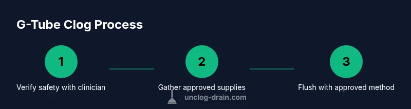
- 1
Confirm safety with clinician
Review whether flushing is appropriate and what method to use. Do not proceed without explicit approval. Ensure all supplies are ready before starting.
Tip: If you are unsure, pause and call your clinician. - 2
Prepare the environment
Wash hands thoroughly, gather supplies, and position the patient for comfort and safety. Ensure the tube is accessible with minimal movement.
Tip: Have a caregiver assist to maintain stability. - 3
Flush using approved method
Attach the syringe to the G-tube port and gently flush with the clinician-approved solution. Do not force the solution if resistance is felt.
Tip: Use slow, steady pressure and pause if resistance increases. - 4
Inspect and reassess
Check for signs of partial relief or continued blockage. If there is no improvement after one or two attempts, stop and contact the care team.
Tip: Document any changes and notify the clinician promptly. - 5
Record and communicate
Document the outcome, the method used, and any symptoms. Inform the care team if fever, vomiting, or leakage occurs.
Tip: Keep a log for future reference. - 6
Follow-up steps
Follow the clinician’s next steps, which may include a re-evaluation, replacement, or different flushing protocol.
Tip: Never perform a replacement or repositioning without professional guidance.
Common Questions
What is a G-tube and why can it become blocked?
A G-tube is a gastrostomy tube that provides access to the stomach for feeding and medications. Blockages occur when thick formulas, medications, or residue accumulate in the tube. Regular flushing per clinician instructions helps prevent this.
A G-tube is a feeding tube through the abdomen. Blockages happen when material builds up; follow your clinician's flushing plan to prevent it.
Can I unclog a G-tube at home?
Only if your clinician has given explicit approval. Do not improvise, and contact the care team for the correct flushing method and any signs to watch.
Only with your clinician’s approval. Don’t improvise and contact your care team for the right method.
What are safe flushing methods?
Safe flushing is determined by your healthcare provider and may involve sterile saline and specific technique. Follow the exact steps they provide and avoid any 'home cures' that aren’t approved.
Flushing methods should be approved by your provider and performed as directed. Do not improvise.
What if flushing doesn’t relieve the blockage?
If flushing fails, contact your care team immediately. Persistent blockage can signal tube issues that require professional assessment or tube replacement.
If flushing doesn’t help, call your care team right away.
How can I prevent future clogs?
Follow a regular flushing schedule and ensure medications are properly dissolved. Work with your clinician to adjust feeds or formulas that reduce residue.
Keep a regular flushing routine and work with your clinician to adjust feeds that reduce buildup.
Watch Video
Key Points
- Always involve a clinician before attempting any unclogging.
- Use only approved flushing methods and solutions.
- Monitor for signs of complications and seek help immediately.
- Prevent blockages with routine, clinician-guided care.